OLYMPUS DSX1000 - Archaeology
We showcased the Olympus DSX1000 to Endinburgh University where Professor Clive Bonsall analysed prehistoric teeth and palaeolithic flint c 30-13000 yr.
''Below are images of two objects in the Archaeology Teaching Collection at Edinburgh University. Firstly we have prehistoric human tooth (molar)
and then a prehistoric flint blade, southwest France, Upper Palaeolithic c. 30-13,000 yr. Image C – magnified view of the enamel on the tooth. The lines look like perykymata, which are natural lines (faint grooves) on the enamel and usually only visible with magnification. They are normal incremental growth lines and should not be confused with dental enamel hypoplasia which refers to (abnormal) defects in the growth of a tooth caused by various factors (malnutrition, disease, etc).
Image E – shows wear polish along the edge of the flint blade resulting from use, possibly cutting through plant stems (I am not a use-wear specialist, so I could be wrong about the material on which the blade was used).
The images illustrate what can be achieved with different settings of the microscope, and why one needs to experiment with the settings''
Professor Clive Bonsall - University of Edinburgh

image a
prehistoric human tooth (molar)

image b
prehistoric human tooth (molar)

image c
prehistoric human tooth (molar)
Prehistoric flint blade, southwest France, Upper Palaeolithic c. 30-13,000 yr.
prehistoric flint blade, southwest France, Upper Palaeolithic c. 30-13,000 yr.

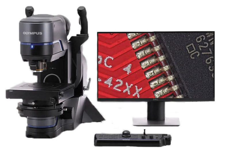

Olympus DSX1000 Digital Microscope all in one
Switch between 6 different observation methods by pushing a button
Fast macro to micro viewing
Accurate measurements with a telecentric optical system


Olympus DSX1000 Digital Microscope all in one
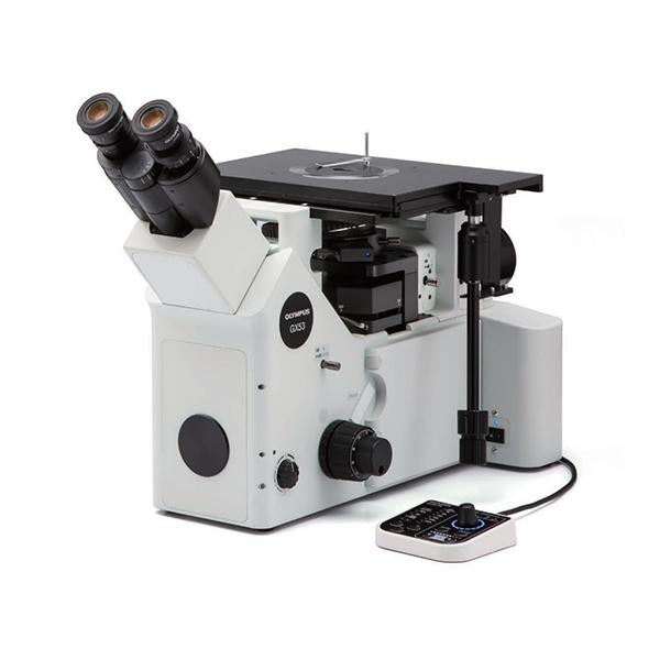


Olympus GX53 Inverted Microscope
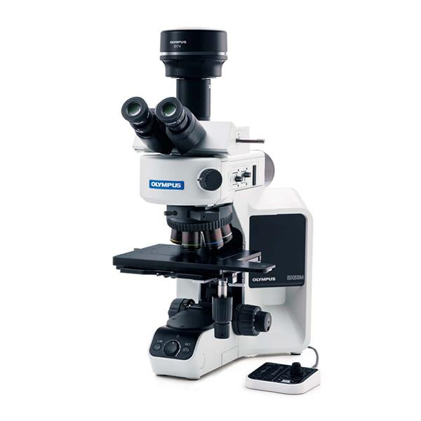

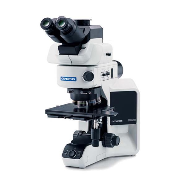
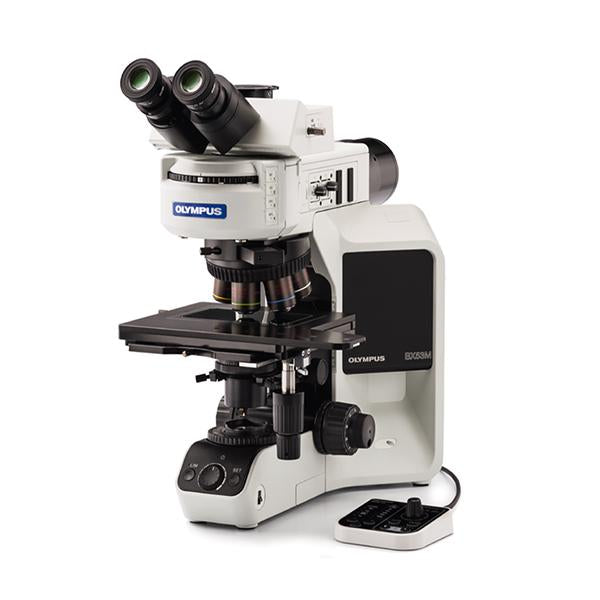
Olympus BX53M Upright Microscope
